Image and Color
An Upside Down Image
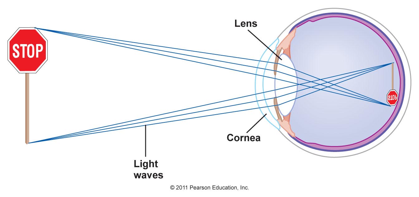
Refraction in the
eye causes light rays from a point on an
object to converge to a focal point on the retina.
Many light rays from many different points on an
object converge on the retina forming an image of
the object.
The image formed on the retina is upside down
(shown in the picture on the right). This is due
to the direction of the light rays as they enter
the eye, and the nature of the converging lens
which do not bend light rays as they enter the
center of the lens.
Retina
The retina
functions as the light detector of the eye.
The retina has two types of nerve
cells that can sense light:
- Rods- located at the
outer edge of the retina. There about 120
million of these cells in the eye and they are
the most sensitive to light, which make them
good motion detectors and for vision in dim
lighting. However they are not sensitive to
different colors.
- Cones- concentrated near
the center of the retina. There are 6 to 7
million of these cells in the eye and they are
responsible for detecting color. The fovea
centralis, located at the very center of
the retina, contains closely packed cones and no
rods, which provide the best resolution and
sensitivity to color.
http://science.howstuffworks.com/life/human-biology/e
ye3.htm
Color
Rods and Cones cells on the retina contain
photosensitive chemicals. The rods contain the
chemical rhodopsin, and the cone contains cone
pigments. There are three types of these pigment:
red, blue, and green. Each cone has one of these
pigments and is therefore sensitive to the
corresponding color.
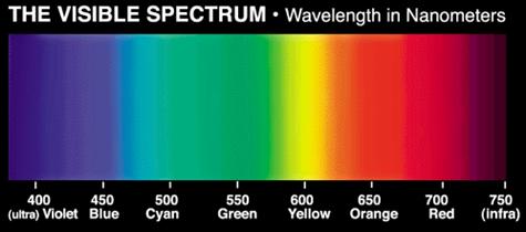
It is important to note that color is a
perception, not a physical entity. The appearance
of color is due to the wavelength of light.
Therefore each cone is sensitive to different
wavelengths of light, and this is then interpreted
by the brain as color. The image to the left shows
the wavelengths of light and their corresponding
color.
http://www.skidmore.edu/~hfoley/Perc7.htm
Brain
Once the light rays produce an image
on the retina, chemical reactions occur at the
rods and cones which then transmits electrical
impulses to the optic nerve. The image is then
sent to part of the brain called the primal
visual cortex which is responsible for
interpreting the image. This is where the image is
flipped right side up.