![]()
Polarizing microscopes are commonly referred to as petrographic microscopes because they are often used to investigate rocks and minerals. Petrographic microscopes use either polarizing sheets or double refractive Nicol prisms to polarize the monochromatic light emerging from the light source. The microscope uses a combination of lenses and mirrors to direct the light from the base upwards towards the stage, a rotating platform upon which thin sections, or microscope slides, are placed. The light is usually diffused and filtered so that it more closely approximates sunlight.
The Lower Polar
On modern microscopes the lower polar is a polarizing sheet consisting of stretched molecules. On earlier microscopes, a Nicol prism was used as the lower polarizer. The lower polarizer usually transmits polarized light virbrating in the front-back direction (or N-S direction).
The petrographic microscope uses two polarizers. The upper polarizer is called the analyzer. The analyzer is mounted so that it can be inserted or removed with ease. It also consists of a polarizing sheet on new microscopes. The vibration directions of the upper and lower polarizers are at right angles to each other. If the lower polarizer transmits light in a N-S orientation, the upper polarizing direction is E-W. When both polars are being used, the polars are "crossed". No light will be transmitted if nothing is on the stage. When the analyzer is not in use, the light seen through the microscope is called "plane light" or "plane polarized light".
Courtesy of Carl Zeiss. Inc. http://www.zeiss.com/
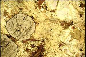
Now that polarization is better understood we can return to the idea of pleochroism. Many minerals show a change in color in plane polarized light as the microscope stage is rotated. This change in color is pleochroism. Pleochroism happens because when light is split into two rays upon entering an anisotropic mieral, the rays of light are absorbed differently as they pass through the mineral.
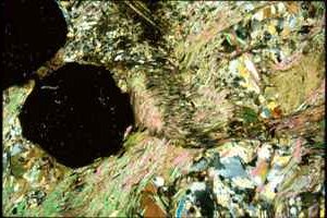
When an anisotropic mineral is looked at through the microscope when the polars are crossed, it is usually light and shows characteristic colors, called interference colors. Interference colors are produced as a result of light being split into two rays upon entering an anisotropic mineral. When the light is split into 2 rays, the two rays have different velocities and indices of refraction. The difference between the index of refraction for the slow and fast rays is called birefringence and is a characteristic property for minerals. Interference colors are produced when the 2 rays are resolved into the vibration direction of the analyzer.
Plane Polarized Light vs. Crossed Polars
Courtesy of New York State Museum http://www.nysm.nysed.gov/splendor_p_pol.html
When the vibration direction of the mineral is oriented parallel to the lower polarizer, the grain looks extinct, or dark, in crossed-polars. Anisotropic minerals go extinct 4 times (every 90o) in 360o rotation of the microscope stage.