Neurons communicate with one
another using two types of electrical signals: graded
potential are used for short distance communication only and
an action potential allows communication over long
distance within the body. A neurons plasma membrane
exhibits a membrane potential, an electrical potential
difference or voltage across the membrane termed the resting
membrane potential. When a neuron is not transmitting an
action potential, the resting membrane potential can be
stated as Vinside
- Voutside
which is typically between -40 mv to -90 mv. A typical
value is -70 mv. The membrane potential is like
voltage stored in a battery. For example, Zoe's car wont
start because her battery is dead and you offer to pass her
current, you connect the cables to the proper terminals and
without really thinking about it, you will have a flow of
electrons and all of the sudden Zoe's car starts. In a
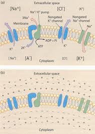
neuron this flow of electric current is composed of ions
such as potassium, sodium and chloride ions that open and
close in response to specific stimuli. Neurons have a
positive charge outside of the cell or extracellular fluid
and a negative charge inside of the cell. The negative
charge inside of the cell is due to the fact that the cells
plasma membrane has more K^+^ ion leakage channels than
Na^+^ leakage channels and therefore potassium can leak out
of the cell easily. As more K^+^ ions leave the
cell, the inside of the cell becomes increasingly negative
and the outside of the cell becomes increasingly positive.
Some of the anions present inside the cell cannot leave the
cell once the K^+^ ion leakage channels are open because
these anions are attached to nondiffusible molecules such as
ADP and large proteins . These trapped anions also
contribute to the resting membrane potential.
that
contributes to the resting membrane potential.
Image retrieved from
http://en.wikipedia.org/wiki/Membrane_potential
Fig 1. Illustrates the
difference in potential across
the plasmamembrane of a neuron and the different ions that cross.
Image retrieved from http://fourier.eng.hmc.edu/e180/lectures/signal1/node3.html