The Imaging Process
Images are actually created using the different frequencies and magnetic fields that are applied to the sample.
Let's sum up all the basics:
*Each voxel has one or two protons, each with a Lamour frequency.
*The Lamour frequency of a proton is based on what type of atom it is and on the strength of the magnetic field acting on it.
*The MRI scans evenly sliced sections, each with a certain number of voxels and protons.
*Each voxel has its own point in the coordinate system of the computer.
*The MRI machine creates a unique magnetic field for each voxel and therefore, a unique Lamour frequency.
*The RF coils in the MRI machine apply RF waves, at a frequency equal to that of the Lamour frequency, to each voxel in the sample.
*When the RF waves are applied to the sample, resonance will occur and the sample will absorb or emit energy and jump from a low energy state to a high energy state and vice versa.
*When the RF waves are turned off, the proton(s) in each voxel will dissipate any absorbed energy and the frequency of this energy is detected and recorded by the computer.
*Each frequency has its own characteristics (i.e. color and size).
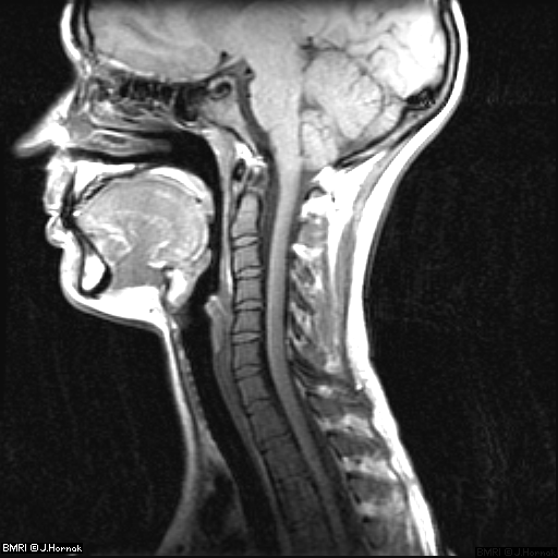
*Essentially, the computer plots each voxel according to its frequency and position (which involves VERY difficult Fourier Transforms) and generates pictures such as this:
This shows a side view of the head and neck. To see more images, click here.
Here are two different views of the same spine. The one on the left is the sagittal view and the one on the right is the axial view.

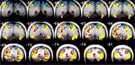
It is also possible to view brain activity with MRI.
The different colors show what level of activity was occurring during a certain task.
Because specialists know what a normal image of a body part looks like, they can compare generated images to these standards.
If there is some type of abnormality in an area of the body, it will be visible in the generated image and a specialized doctor will interpret the results.